Action potential
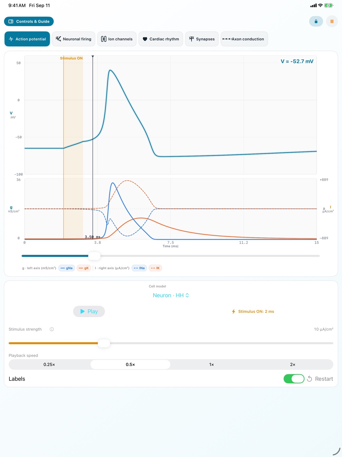
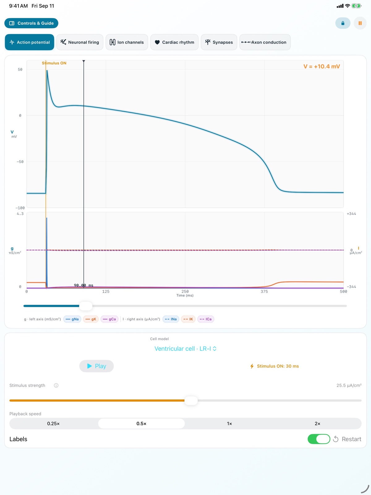
Explore Hodgkin–Huxley neuronal and Luo–Rudy I ventricular-cell models. Move the time cursor to revisit particle positions, gates, g, I, and V.
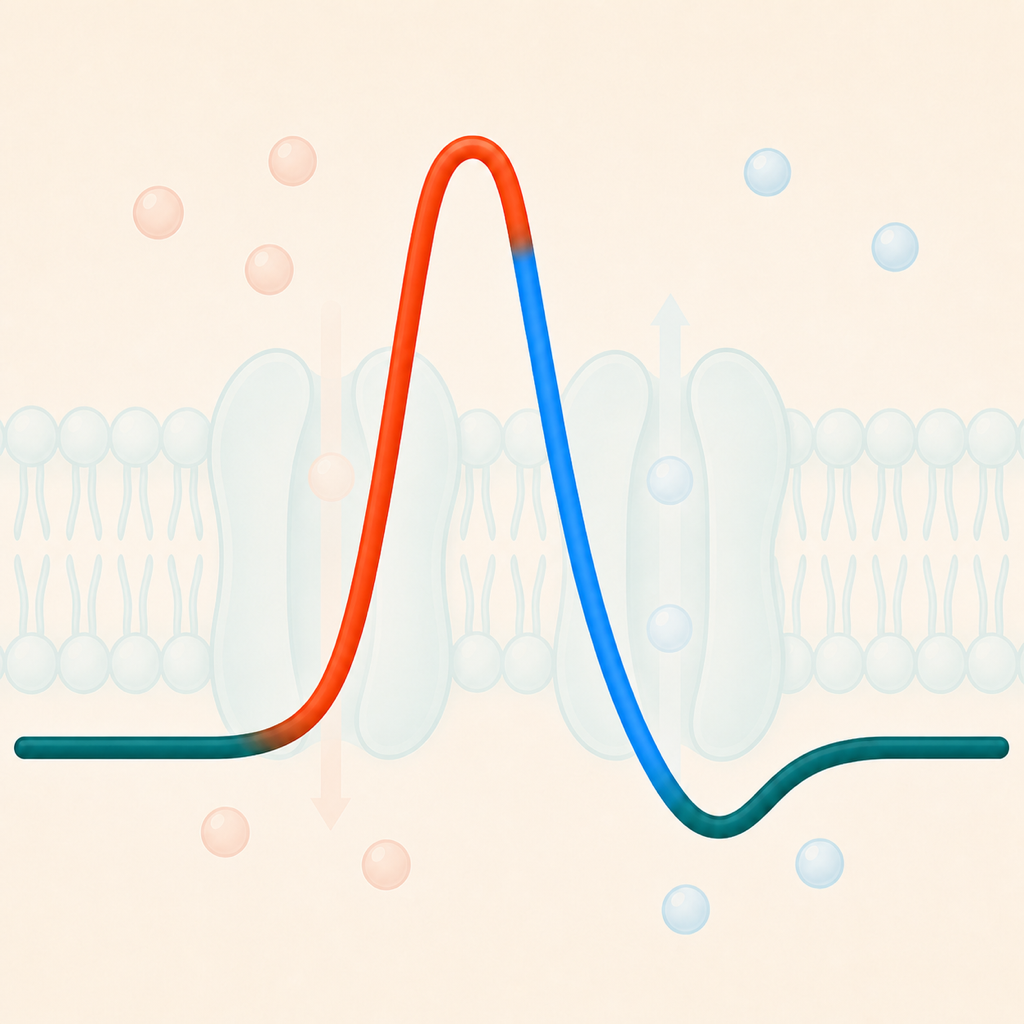
Connect membrane voltage with ion movement and waveforms.
See ion particles, channel gates, conductance, current, and membrane voltage at the same model time. Follow excitable-cell behavior from neuronal and ventricular action potentials to neuronal firing, cardiac conduction, synaptic integration, and axon propagation. In each simulation, adjust parameters suited to the model—such as stimulus strength and duration, channel conductance, or input conditions—and compare how the waveform or propagation changes.
Six simulations
Observe the same physiology at different scales, with in-app guidance on what to look for in each trace.
Explore Hodgkin–Huxley neuronal and Luo–Rudy I ventricular-cell models. Move the time cursor to revisit particle positions, gates, g, I, and V.
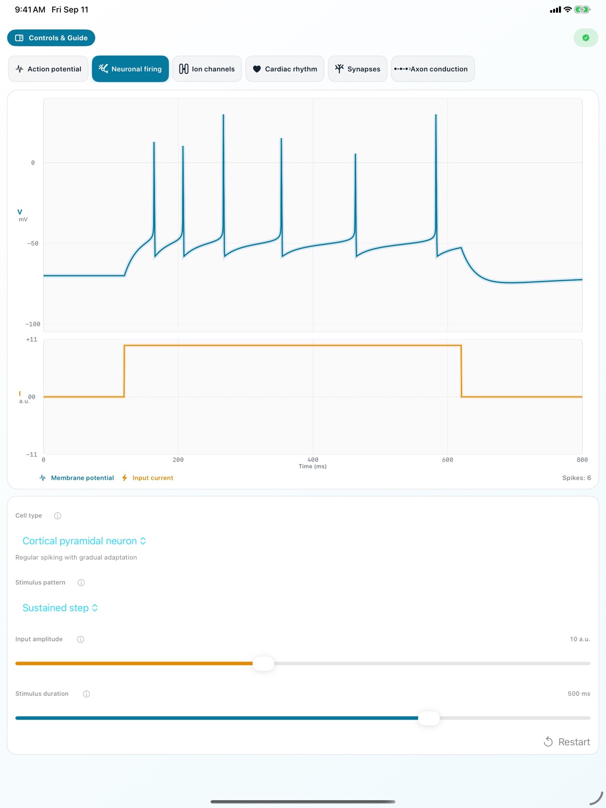
Compare nine profiles: cortical pyramidal, fast-spiking interneuron, medium spiny, cholinergic, hippocampal CA1, Purkinje, thalamic relay and reticular, and midbrain dopaminergic.
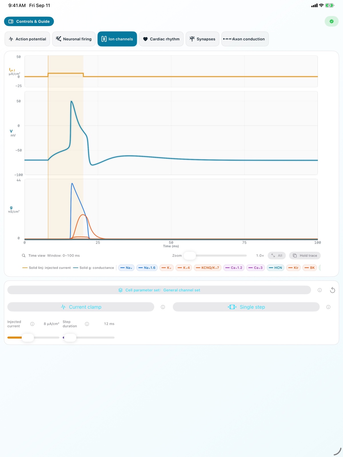
Separate 13 channel types under current or voltage clamp. Adjust the cell set, stimulus, pulse length, channel availability, and maximum conductance.
Follow conduction from sinus node to ventricles, then compare normal rhythm, sinus bradycardia or tachycardia, first-degree AV block, and 2:1 second-degree AV block.
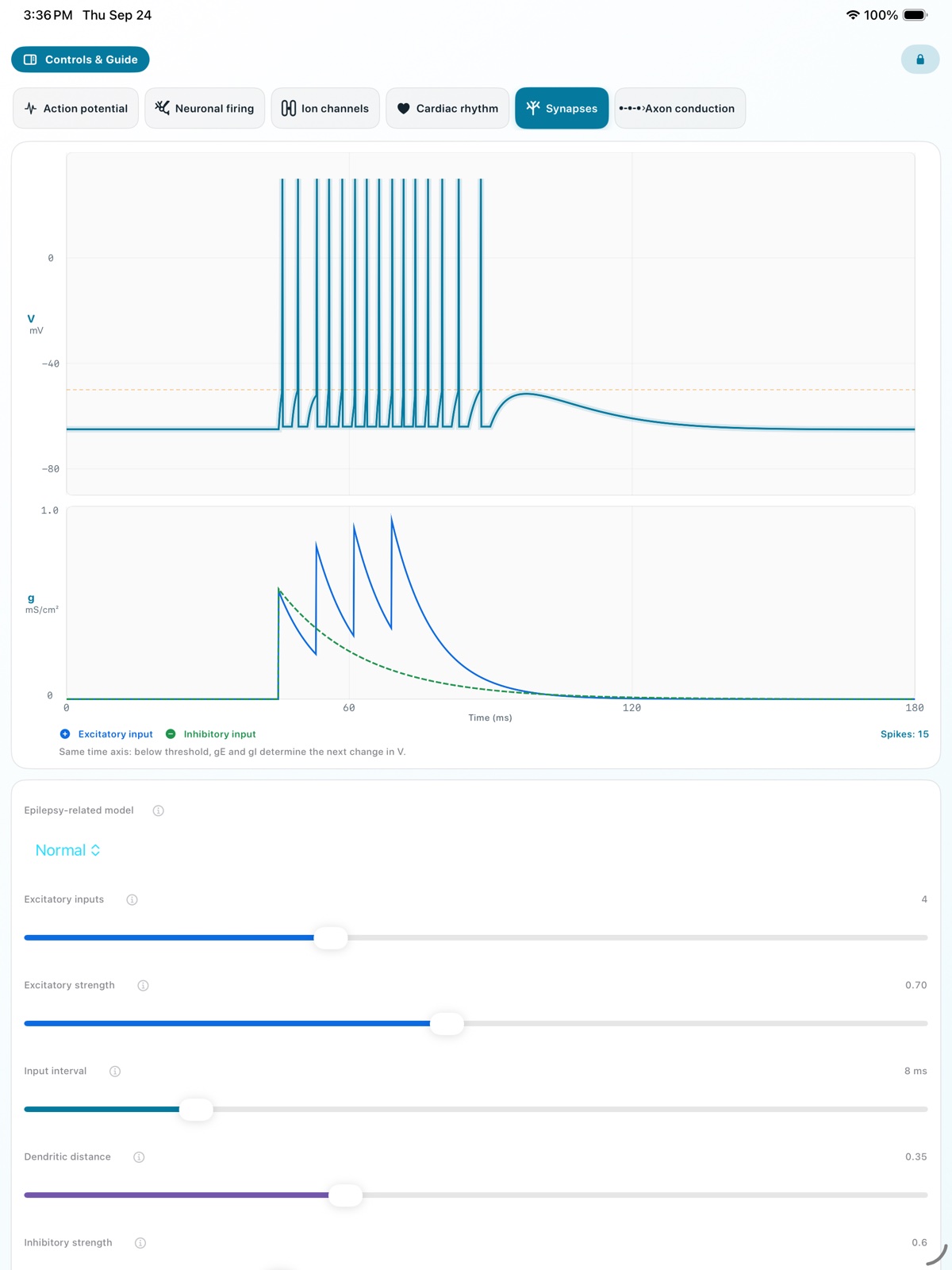
Relate excitatory and inhibitory conductance (gE and gI) to membrane voltage and firing. Compare normal activity with increased excitation or reduced inhibition.
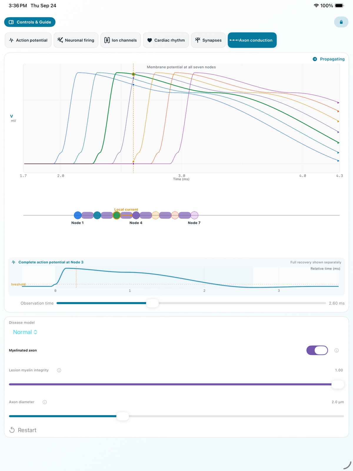
Match the axon diagram to nodal waveforms. Compare normal propagation with focal demyelination, including current leakage, safety factor, subthreshold depolarization, and conduction failure.
What to observe
Use one time cursor to see fast Na⁺ conductance rise before rapid depolarization, followed by delayed K⁺ conductance that drives repolarization and transient afterhyperpolarization.
Compare spike-frequency adaptation, sustained fast firing, delayed first spikes, and post-stimulus rebound. Each cell panel states the feature and the point to inspect.
Hold and overlay a standard trace or switch between normal and disease-related conditions to find where amplitude, timing, or propagation first diverges.
Read injected current, voltage command, or synaptic input on its own plot, then connect that input to changes in conductance and membrane voltage.
In the app
Every image below is an actual iPad screen. See what each simulation reveals, from whole-cell action potentials and individual ion channels to circuits and propagation along an axon. Change the parameters freely and test how the response changes under different conditions.
Included
Use every simulation, playback speeds, the time cursor, observation guidance, normal and disease-related conditions, basic parameters, and four foundational neuron presets.
Pro
Unlock result capture across all six simulations, latest-two numerical comparison, CSV export, advanced Action Potential controls, held Ion Channel traces, and five specialized neuron presets. This is not a subscription. Other storefronts use the corresponding price shown by the App Store.
Requirements & privacy
Requires iOS/iPadOS 17 or later or macOS 14 or later. The interface supports English, Japanese, Korean, Simplified and Traditional Chinese, Spanish, French, German, Italian, and Brazilian Portuguese.
The App has no user accounts, ads, or analytics SDKs. The App is for learning and visualization, is not a medical device, and must not be used for diagnosis or treatment.